
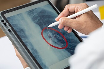
This year marks 125 years since the accidental discovery of the X-ray, when Wilhelm Rontgen, a German physicist, successfully produced a ray that was capable of passing through most substances. In the years after the discovery, doctors first used X-rays to diagnose gunshot wounds and bone fractures. While Rontgen went on to earn the first Nobel Prize in physics for his efforts in 1901, more than a century later, the potential for X-rays keeps growing, even during a global pandemic.
Recent medical news reveals that X-rays are at the forefront of diagnostic tools used to identify lung abnormalities related to COVID-19. X-rays may also be part of the medical innovative solutions that help patients recover. In August 2020, the National Institutes of Health (NIH) announced the launch of the Medical Imaging and Data Resource Center, which will use artificial intelligence and medical imaging to accomplish early detection and personalized therapies for COVID-19 patients.
The scope of X-rays encompasses different diagnostic procedures used for years, including arteriograms to examine arteries, computed tomography (CT) scans, and fluoroscopy, the study of moving body structures, which is similar to an X-ray "movie." A number of standards support X-rays to ensure the safety of people who depend on them. In the years after the accidental X-ray discovery, physicists and physicians alike were unaware that large X-ray radiation doses could cause serious, lasting effects on the human body. During that time, people also lacked the proper instruments to measure the strength of the radiation fields.
A number of standards set guidelines to help support safer X-rays and medical imaging.
From Broken Bones to Disease Diagnosis: Medical Imaging is Essential to the Healthcare Field
ASTM International, an ANSI member and accredited standards developer, has developed several standards that address X-rays and CT technology. ASTM E1742 / E1742M, Standard Practice for Radiographic Examination, establishes the minimum requirements for radiographic examination for metallic and nonmetallic materials.
Another ASTM standard supports safer CT scans, which are used to see detailed images of the body and to help diagnose muscle and bone disorders. ASTM E1570-19, Standard Practice for Fan Beam Computed Tomographic (CT) Examination, establishes the minimum requirements for computed tomography (CT) examination of test objects using fan beam systems.
The International Electrotechnical Commission, (IEC) has developed the standard IEC 61267 Ed. 2.0 b: 2005, Medical Diagnostic X-Ray Equipment - Radiation Conditions For Use In The Determination Of Characteristics, which applies to test procedures which, for the determination of characteristics of systems or components of medical diagnostic X-ray equipment, require well-defined radiation conditions. Via the U.S. National Committee, ANSI is the U.S. representative the International Electrotechnical Commission (IEC).
Another standard that supports safer medical imaging is ISO 12052:2017, Health informatics - Digital imaging and communication in medicine (DICOM) including workflow and data management. Published by the International Organization for Standardization (ISO), the standard addresses the exchange of digital images and information related to the production and management of those images, between both medical imaging equipment and systems concerned with the management and communication of that information.
The document was developed by ISO Technical Committee (TC) 215, Health informatics. ANSI administers the U.S. Technical Advisory Group (TAG) to ISO TC 215 to coordinate national standards activities for existing and emerging health sectors. The U.S. TAG is guided by the ANSI cardinal principles of consensus, due process, and openness. Read more about ISO TC 15's efforts to support health in a related ANSI article, ISO TC 215 Health Informatics Develops Technical Report on Medicinal Products.
Security Personnel Need a Safety Barrier: X-Rays Get Standards' Support
While X-rays are an essential tool across the medical field, they're also an integral part of airport security, for studying archeology and astronomical objects, and even detecting counterfeit artwork.
"People screening" at airports, prisons, courthouses, and some museums are a common practice in order to detect concealed explosives or other weapons. Standards help to assure better safety for all.
ANSI organizational member, Health Physics Society (HPS) has published radiation protection standards, including ANSI/HPS N43.17,Radiation Safety for Personnel Security Screening Systems Using X-Ray or Gamma Radiation. Two Accredited Standards Committees (ASC)—N13 and N43—along with the HPS standards coordinator, constitute the HPS standards organization.
IEEE, an ANSI member and accredited standards developer, has developed IEEE N42.47-2010, American National Standard for Measuring the Imaging Performance of X-Ray and Gamma-Ray Systems for Security Screening of Humans. The standard applies to security screening systems that utilize X-ray or gamma radiation and are used to inspect people who are not inside vehicles, containers, or enclosures. Specifically, this standard applies to systems used to detect objects carried on or within the body of the individual being exposed. The purpose of this standard is to provide standard methods of measuring and reporting imaging quality characteristics and establish minimum acceptable performance requirements.
These are just a sampling of the many standards and standards developing organizations that support medical imaging. For a full scope of X-ray standards, visit ANSI.org.